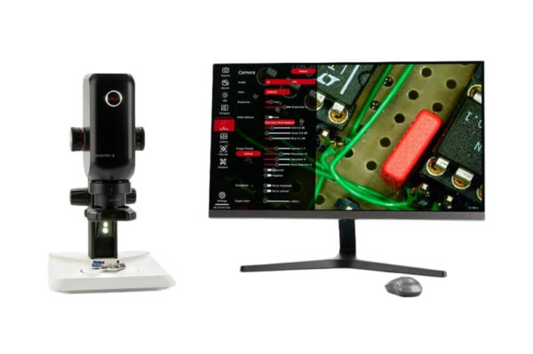
Leica – Emspira 3 Digital Microscope
The advantage of optimized inspection and adaptability to your needs combined in a single system is waiting for you. Save time when meeting demanding throughput targets and inspecting diverse samples with Emspira 3.
For efficient decision making, benefit from secure storage and easy sharing of documentation – no matter if you inspect in stand-alone mode or with a PC. The robust design of Emspira 3 allows you to focus confidently and reliably on your inspection work both in production and laboratory environments.
Technical Specifications
| General optical data | |
| Max. resolution | 337 lp/mm |
|---|---|
| Max. FoVx | 76,1 mm |
| Max. FoVy | 42,8 mm |
| Max. DoF | 40,5 mm |
| Working distance | 303 – 19 mm |
| Optical data for PlanApo 1.0x | |
| Max. measurement 1.0x accuracy @ 0,75x zoom | +/- 1% |
| Max. measurement accuracy @ 6.0x zoom | +/- 0.5% |
| Objective optics carrier | |
| Coding | Click-stop feature |
| Click-stop feature | Eight switchable positions, for repetitive tasks |
| Microscope camera specifications | |
| Live image on an HDMI monitor | at up to 60 fps (3,840 × 2,160 pixels) |
| Full-screen image capture | at 12 MP |
| File formats | JPG, TIF, BMP |
| Housing | |
| Antimicrobial surface | AgTreatTM according to ISO 22196 |
| Protection rating | IP 21 |
| Magnification range | |
| Monitor 10″ with Achr. objective 0.32x | total magnification 23:1 |
| Monitor 28″ with Achr. objective 0.32x | total magnification 65:1 |
| Monitor 10″ with PlanApo objective 5.0x | total magnification 360:1 |
| Monitor 28″ with PlanApo objective 5.0x | total magnification 1027:1 |
| Software & connectivity | |
| Compatibility | USB 3.0, standard USB type C, lockable |
| High-definition connector | HDMI 2.0a, HDMI plug type A |
| USB connectors | 4x USB 2 connectors, type A |
| Controls | USB mouse Optional: keyboard, touch screen, footswitch |
| Supported software | LAS X 5.0.2 and higher |
Key Features
Measure directly during visual inspection without a PC
In stand-alone mode, measure multiple regions on the sample in the live image
Directly compare to references with a single click
For easier decisions, compare the live image to reference images or customized overlays to check if parts are within tolerances.
Annotate images without a PC
- Add annotations directly to the image using the integrated on-screen display
- Easily highlight features and areas of interest on the sample by adding comments and conclusions, as text and graphical elements, to the image
Automatically save images on your network
- Save images and results from inspection directly to your local network via ethernet connectivity for fast storage
- Simultaneously take and share images with a single press of a button using the optional hand/foot switch
Share results via email with one click
Easily share documentation directly with email contacts without a PC
Use the illumination that works for your sample
Reveal relevant details with the appropriate illumination
How It Works
Leica – Emspira 3 Digital Microscope
- Digital microscope
- Quick sample identification
- PCB Inspection
- On-screen display
Emspira 3 Digital Microscope
The Emspira 3 digital microscope empowers you to streamline your inspection process, cover your needs flexibly, and work with confidence. It combines everything needed to perform comprehensive visual inspection with a single system, including comparison, measurement, and data sharing.
Emspira 3 – Inspect With a Lean Setup
Emspira 3 combines everything needed to perform comprehensive visual inspection into a single system, including comparison, measurement, and documentation sharing. Inspect with a single system – no need for a PC. Streamline your inspection process with a lean.
More Products
Leica Microsystems develops and manufactures microscopes and scientific instruments for the analysis of microstructures and nanostructures. Widely recognized for optical…


